

The Department of Earth Sciences Electron Probe Microanalyser (EPMA) Laboratory is a departmental facility that offers analytical training and services to members of the Department of Earth Sciences, other university researchers and some commercial clients.
Booking the equipment and receiving appropriate training before using the microprobe is mandatory.
Use the table below to learn about the equipment and services available in the lab and to view the booking calendar. Please also check out the gallery tab to see pictures of recent application examples.
Need to prepare your samples before analysis? Click the button below for details on sample preparation:
Fill the ![]() Job Request Form to proceed: contact the technician for questions.
Job Request Form to proceed: contact the technician for questions.
For more information, contact the Microprobe Technician:
Dr. Yanan Liu
Lab Manager (Microprobe/SEM/XRD Labs)
Phone: 416-978-5420
Email Dr. Liu
JXA-8230 Key Product Features (from JEOL USA)
| Detectable element range | 4Be to 92U |
| Detectable wavelength range | 0.087 to 9.3 nm |
| Number of X-ray spectrometers 5 (Plus EDS) | 5 (Plus EDS) |
| Specimen size | 150 mm x 150 mm x 50 mm |
| X – Y Range | 90 mm x 90 mm |
| Specimen stage drive speed | 15 mm/s max |
| Accelerating voltage | 0.2 to 30 kV (100 V steps) |
| Probe current range | 10-12 to 10-5 A |
| Probe current stability | 1 x 10⁻³/h |
| BEI (backscattered electron image) | TOPO and COMPO |
| Scanning image magnification | 40x to 300,000x (WD: 11 mm) |
Exciting new features, including but not limited to:
- 5 WDS, which would cut the counting time into halves;
- Improved EDS system detecting B~U;
- Fully integrated WDS and EDS system, which enables major elements measurements by EDS, trace by WDS to save even more time;
- Integrated imaging package, which will better record the probed locations and make “mapping with WDS” become possible;
- Optional Xenon detector and special spectrometer with smaller Rowland circle which enhances the count rates for metals;
- Stage holder with greater capacities and tolerance
For long distance users, you do not have to come to the lab to set up your probe points.
Probe users can now mail their samples to us and pick your points remotely at home! (Windows system only)
Contact the probe technician about more details!
Our probe lab has been recently upgraded with a new JEOL JXA8230 5-WDS electron microprobe.
Electron Probe X-ray Microanalyser
- The Electron Probe X-ray Microanalyser (EPMA) is used to determine the chemical composition of solid materials on a microscopic scale, down to volumes of a few cubic micrometers.
- In this analytical technique a very narrow beam of high-energy electrons is focused at a selected point on the surface of a flat and highly polished sample. As the electrons penetrate the sample, their energy is first released into and then re-emitted from the sample, via different processes giving rise to different types of signals, each of which carries information about some property of the sample.
X-ray signal
- The X-ray signal carries information about the chemical composition of the micro-volume where it was generated. By means of an appropriate detection system, it is possible to eventually obtain qualitative (elements present) and quantitative (weight %) analyses. The detection of the X-rays is accomplished by means of crystal spectrometers (WDS, wavelength dispersive spectrometry) and solid state Si(Li) detectors (EDS, energy dispersive spectrometry). A computer and specialized software are also needed to achieve that end, ultimately making it possible to generate large volumes of high quality data, either in a manual or in an automatic, unattended mode.
Other signals
- Other signals typically utilised in an electron microprobe are Secondary Electrons (SE) and Backscattered Electrons (BSE). The SE emission intensity is most strongly modulated by surface topography, that of BSE by compositional variations. Both signals are collected while the beam is being scanned on a very small portion of the sample surface. The reconstruction of the spatial distribution of the collected signal intensity generates two-dimensional images displaying surface relief (SE) and compositional contrast (BSE).
Our Equipment
- Our microprobe is a JEOL JXA8230 instrument, with 5 wavelength dispersive spectrometers and various combinations of different diffracting crystals:
| Crystal | (A) | Range of elements |
|---|---|---|
| TAP | 25.757 | ~ P (Ka ) |
| LIF, LIFH | 4.027 | ~ Rb (Ka) |
| PETJ, PETH | 8.742 | Si ~ Mn (Ka) |
| LDE1 | 60 | ~ F (Ka) |
| LDE2 | 95 | ~ N (Ka) |
Detection Limits depends on the element of interest and its matrix, (e.g. 6 ppm for Fe, <30 ppm for Se, etc.). Detection limit in weight percent for a single analysis line with a confidence of 99% (assumes 3 standard deviations above the background, JEOL).
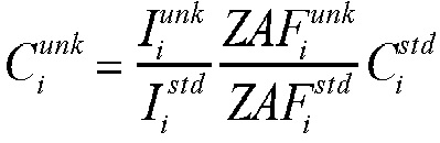
Where:
ZAF is the ZAF correction factor for the sample matrix
I is the raw count rate on the analytical standard and unknown samples
Cstd is the pre-determined concentration in the standard
Spatial resolution varies from ~3 microns in diameter for small particles to mm2 level for raster areas.
Penetrating depth can be varied for film/coating analyses, varying from submicron to over 10 um depending on the element of interest and its matrix.
Liquid nitrogen free, high count-rate silicon drift detector (SDD) EDS spectrometer.
Fees
The current fee structure (in $ per hour) is found below. Note that training will need to be arranged separately at a cost (contact the lab manager for details).
| Users | Peak hours (9 am - 5 pm weekdays)2 | Off-peak hours (5 pm - 9 am, weekends and holidays)2 |
|---|---|---|
| Preferred users (contribute $2,500 upfront)1 | $45 / hour | $35 / hour |
| Academic regular users (including other departments and other universities) | $60 / hour | $50 / hour |
| Mapping (for academic users) | $35 / hour | $35 / hour |
| Commercial and contract users | $120 / hour | $80 / hour |
| Mapping (for commercial and contract users) | $60 / hour | $60 / hour |
1Users can contribute upfront $2,500 will get the above discounted rate. The user fees will be offset against this amount ($2,500). If the $2,500 is not used up during the fiscal year, the remaining amount will be rolled over to the next year at regular rate.
2An additional 1 hour setup time will be automatically charged for every analytical session.
Important notes for booking:
- Every calendar week, each user may only book 1 day for on-peak time use, so that everyone gets to use the machine in a timely manner.
- Off-peak time can only be booked by independent users. Please consult lab personnel for availability.
- Cancellations made 24 hours or less before an appointment will be subject to a two hours flat rate charge.
- In special circumstances, the management may take over already booked time slots for necessary maintenance and emergencies. Alternative booking options will be discussed.
- Booking is arranged based on the time sequence of the booking request in most cases; in some special cases, priority may be given to users from a long distance away (i.e., another city) or to those who have specified deadlines (within one week).
- Undergraduate students (departmental) need to fill out a special job request form (found here) with supervisor’s signature, in order to get a maximum 50 hours discount on the 4th year thesis project only. Ask the technician for more info.
- Alternative arrangement may be agreed upon by users, provided that everyone’s rights are respected.
EPMA mapping example: zoning in chondrites (courtesy of Royal Ontario Museum (ROM), 2021)
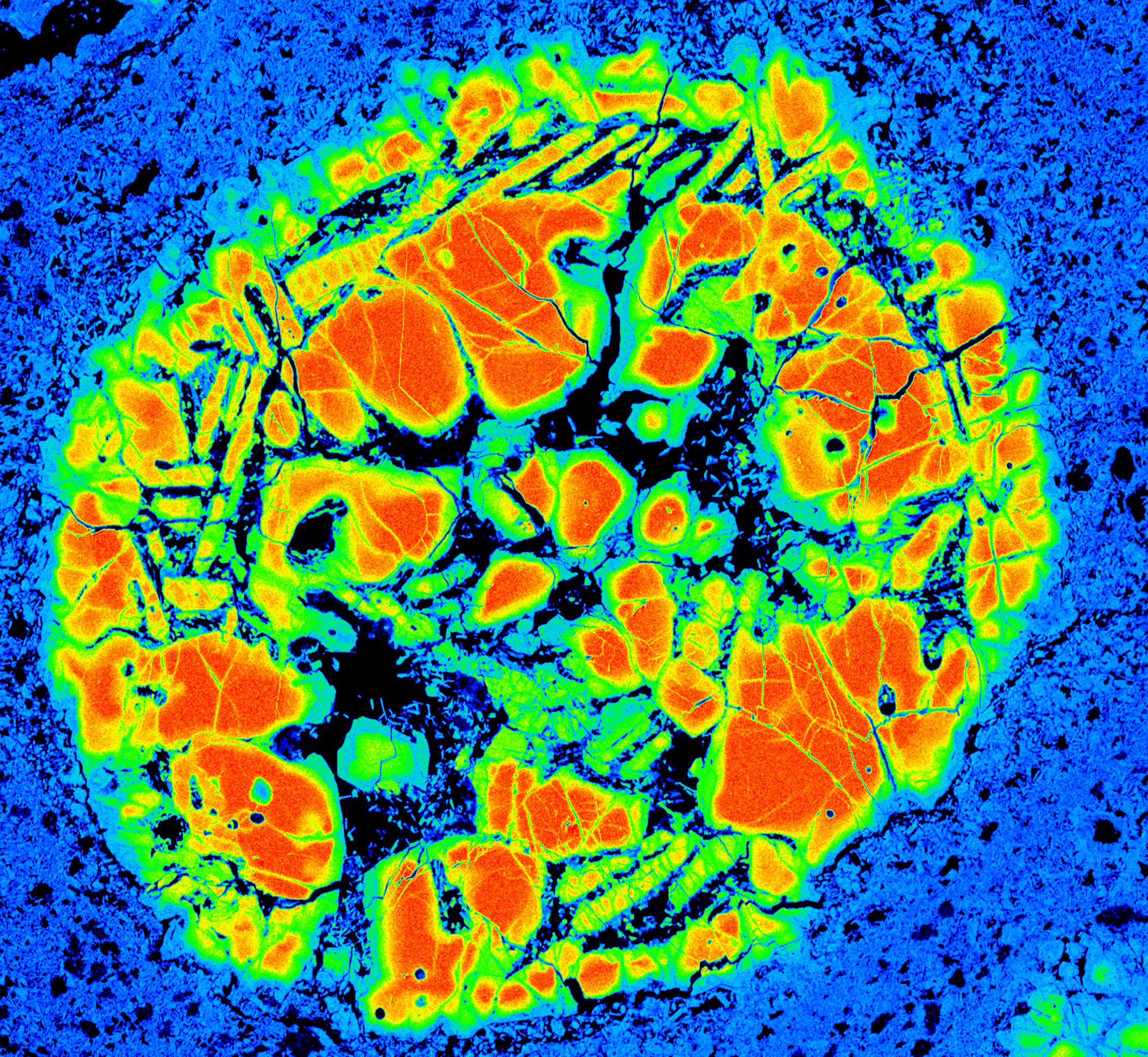
View more photos in the gallery.